


The main treatment method for varicose veins (VV) remains surgery. The goal of the operation is to eliminate the symptoms of the disease (including cosmetic defects) and prevent the progression of varicose transformation of the saphenous veins. Today, none of the existing surgical methods alone meets all the pathogenetic principles of treatment, as a result, the need for their combination becomes obvious. Various combinations of certain operations depend mainly on the severity of pathological changes in the venous system of the lower extremities.
The indication for surgery is the presence of blood reflux from the deep veins to the superficial veins in patients with classes C2-C6. A combined operation may include the following steps:
- Ligation of the estuary and intersection of the GSV and/or SVC with all tributaries (crossectomy);
- Removal of GSV and/or SSV trunks;
- Removal of varicose tributaries of the GSV and SSV;
- Crossing of incompetent perforating veins.
This area of operation has been developed over decades of scientific and practical research.
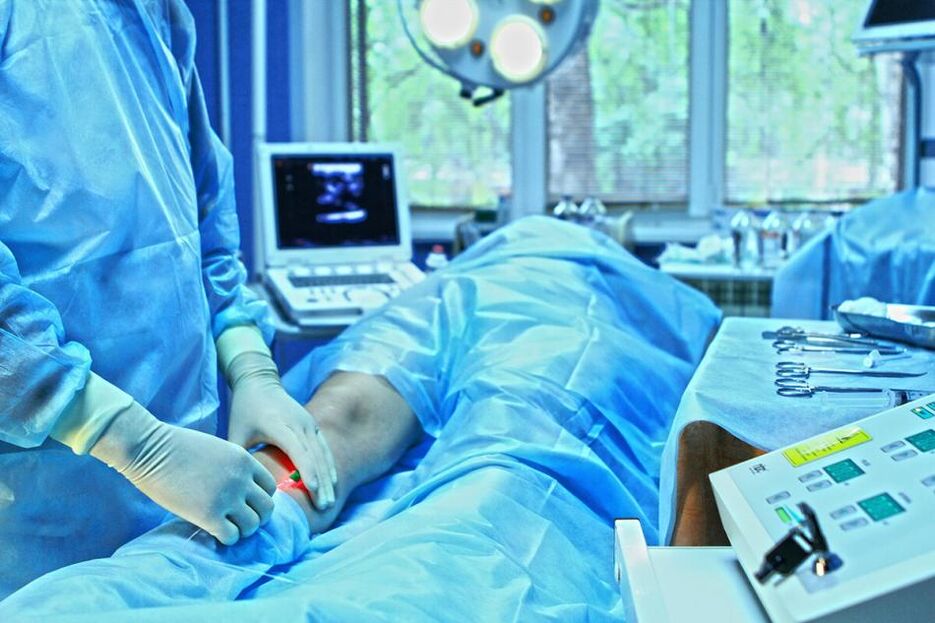
Crossectomy of the great saphenous vein. The optimal approach to ligate the VGS is through the inguinal fold. The suprapinguinal approach has some advantages only in patients with recurrent disease due to the remaining pathological stump of the GSV and a high location of the postoperative scar. The GSV must be ligated strictly parietal to the femoral vein; All estuarine tributaries must be ligated, including the upper one (superficial epigastric vein). There is no need to suture the oval window or subcutaneous tissue after GSV crossectomy.
Removal of the trunk of the great saphenous vein. When determining the extent of removal of the GSV, it is necessary to take into account that in the vast majority of cases (80-90%), reflux along the GSV is recorded only from the mouth to the upper third of the Leg. Removal of the GSV along its entire length (full extraction) is accompanied by a significantly higher incidence of damage to the saphenous nerves compared to removal of the GSV from the mouth to the upper third of the leg (short extraction): 39 % and 6. 5%, respectively. At the same time, the frequency of relapses of varicose veins does not differ significantly. The remaining segment of the vein can be used in the future for reconstructive vascular operations.
In this sense, the basis of intervention in the GSV basin should be short clearing. Removal of the entire length of the trunk is permitted only if it has been reliably confirmed to be incompetent and has expanded significantly (more than 6 mm in a horizontal position).
When choosing a saphenectomy method, preference should be given to intussusception techniques (including PIN removal) or cryoflebectomy. Although detailed studies of these methods are still being carried out, their advantages (less traumatic) compared to the classic Babcock technique are undoubted. However, the Babcock method is effective and can be used in clinical practice, but it is advisable to use small diameter olives. When choosing the direction of vein extraction, preference should be given to traction from top to bottom, that is, retrograde, with the exception of cryoflebectomy, the technique of which involves antegrade extraction of the vein.
Crossectomy of the small saphenous vein. The structure of the terminal section of the small saphenous vein is very variable. As a rule, the SVC fuses with the popliteal vein a few centimeters above the line of knee flexion. In this sense, the approach for SVC crossectomy should be moved proximally, taking into account the location of the saphenopopliteal anastomosis (before the operation, the location of the anastomosis should be clarified by ultrasound).
Removal of the trunk of the small saphenous vein. As with GSV, the vein should be removed only to the extent that reflux is determined to occur. In the lower third of the leg, reflux along the SVC is very rare. Invagination methods should also be used. Cryoplebectomy of the SVC has no advantages over these techniques.
A comment. The intervention on the small saphenous vein (crossectomy and removal of the trunk) must be performed with the patient in a prone position.
Thermoobliteration of the main saphenous veins. Modern endovasal techniques (laser and radiofrequency) can eliminate brainstem reflux and therefore, in terms of their functional effect, can be considered an alternative to crostectomy and extraction. The morbidity of thermoobliteration is significantly less than that of stem phlebectomy and the cosmetic outcome is significantly greater. Laser and radiofrequency obliteration is performed without ostial ligation (GSV and SSV). Simultaneous crossectomy virtually eliminates the benefits of thermoobliteration and increases the cost of treatment.
Obliteration with endovasal laser and radiofrequency has limitations in its use, is accompanied by specific complications, is much more expensive and requires mandatory intraoperative ultrasound control. The reproducibility of the technique is low, so it should only be performed by experienced specialists. The long-term results of its use in widespread clinical practice are still unknown. In this sense, thermoobliteration methods require further study and cannot yet completely replace traditional surgical interventions for varicose veins.
Removal of varicose veins. When removing varicose tributaries from the superficial trunks, preference should be given to their removal using mini-phlebectomy instruments using skin punctures. All other surgical methods are more traumatic and lead to worse cosmetic results. According to the patient, it is possible to leave some varicose veins, which are subsequently eliminated through sclerotherapy.
Dissection of perforator veins. The main controversial issue in this subsection is the determination of the indications for intervention, since it is necessary to clarify the role of perforators in the development of chronic venous disease and its complications. The inconsistency of numerous studies in this area is associated with the lack of clear criteria for determining the incompetence of perforator veins. Several authors generally question the fact that incompetent perforating veins may have an independent significance in the development of CVD and be a source of pathological reflux from the deep to the superficial venous system. The main role in varicose veins is assigned to vertical discharge through the saphenous veins, and the failure of perforators is associated with the increasing load on them to drain reflux blood from the superficial to the deep venous system. As a result, they increase in diameter and have bidirectional blood flow (mainly into the deep veins), which is determined primarily by the severity of the vertical reflux. It should be noted that bidirectional blood flow through perforators is also observed in healthy people without signs of CVD. The number of incompetent perforator veins is directly related to the CEAP clinical class. These data are partially confirmed by studies in which, after interventions in the superficial venous system and elimination of reflux, a significant proportion of perforators become solvent.
However, in patients with trophic disorders, 25. 5% to 40% of perforators remain incompetent and their further impact on the course of the disease is unclear. Apparently, with varicose veins of classes C4-C6 after elimination of vertical reflux, the chances of restoring normal hemodynamics in perforator veins are limited. As a result of prolonged exposure to pathological reflux of subcutaneous and/or deep veins, irreversible changes occur in a certain part of these vessels and the reverse flow of blood through them acquires pathological significance.
Therefore, today we can talk about mandatory careful ligation of incompetent perforator veins only in patients with varicose veins with trophic disorders (classes C4-C6). In clinical classes C2-C3, the decision on ligation of perforators should be made individually by the surgeon, depending on the clinical picture and data from instrumental examination. In this case, dissection should only be performed if its failure is reliably confirmed.
If the location of trophic disorders excludes the possibility of direct percutaneous access to an incompetent perforator vein, the operation of choice is endoscopic subfascial dissection of perforator veins (ESDPV). Numerous studies indicate its undeniable advantages compared to the previously widely used open subfascial subtotal ligation of perforators (Linton operation). The incidence of wound complications with ESDPV is 6-7%, while with open surgery it reaches 53%. At the same time, the healing time of trophic ulcers, indicators of venous hemodynamics and the frequency of relapses are comparable.
A comment. Numerous studies indicate that ESDPV can have a positive effect on the course of chronic venous disease, especially when it comes to trophic disorders. However, it is not clear which of the observed effects are due to dissection and which to concurrent saphenous vein surgery in most patients. However, the lack of long-term results in patients with C4-C6 who did not undergo interventions on perforator veins, but only phlebectomy, does not yet allow us to draw final conclusions about the use of certain methods of surgical treatment.
Despite the existing contradictions, most researchers still consider it necessary to combine traditional interventions on superficial veins with ESDPV in patients with trophic disorders and open trophic ulcers against the background of varicose veins. The ulcer recurrence rate after phlebectomy combined with ESDPV ranges from 4% to 18% (follow-up period 5 to 9 years). In this case, complete healing occurs in approximately 90% of patients within the first 10 months.
Good results were also obtained when other minimally invasive techniques were used to remove perforator veins, such as microfoam scleroobliteration and endovasal laser obliteration. However, the likelihood of success with their use directly depends on the qualifications and experience of the doctor, so they cannot be recommended for widespread use for now.
In patients with clinical classes C2-C3, ESDPV should not be used, since removal of perforator reflux can be successfully performed from small incisions (up to 1 cm) and even from skin punctures using mini-phlebectomy instruments. .
Correction of deep venous valves. Currently, in this section of surgical phlebology there are more questions than answers. This is due to existing contradictions regarding aspects such as the importance of deep venous reflux and its impact on the course of CVI, the determination of indications for correction, and the evaluation of treatment effectiveness. Failure of various segments of the deep venous system of the lower extremities leads to various hemodynamic disorders, which is important to take into account when choosing a treatment method. Several studies indicate that reflux through the femoral vein does not play any important role. At the same time, damage to the deep veins of the leg can lead to irreparable changes in the functioning of the musculovenous pump and severe forms of CVI. It is difficult to evaluate the positive effects of correction of venous reflux in deep veins, since these interventions in most cases are performed in combination with operations on superficial and perforating veins. Isolated elimination of reflux through the femoral vein does not affect venous hemodynamics at all or causes minor temporary changes only in some parameters. On the other hand, only the elimination of reflux along the GSV in varicose veins in combination with insufficiency of the femoral vein leads to the restoration of valve function in this venous segment.
Surgical methods for treating primary deep venous reflux can be divided into two groups. The first involves phlebotomy and includes internal valvuloplasty, transposition, autotransplantation, creation of new valves, and the use of cryopreserved allografts. The second group does not require phlebotomy and includes extravasal interventions, external valvuloplasty (transmural or transcommissural), angioscopy-assisted extravasal valvuloplasty, and percutaneous installation of corrective devices.
The question of correction of deep venous valves should be raised only in patients with recurrent or non-healing trophic ulcers (class C6), mainly with recurrent trophic ulcers and reflux in deep veins of grade 3-4 (up to the level of the knee). joint) according to Kistner's classification. If conservative treatment is ineffective in young people who do not wish to be prescribed lifelong compression stockings, surgery can be performed for severe edema and C4b. The decision to operate should be made based on the clinical state, but not on data from special studies, since symptoms may not correlate with laboratory parameters. Surgeries to correct deep venous valves should only be performed in specialized centers with experience in this type of intervention.
Surgical treatment of postthrombotic disease.
The results of surgical treatment of patients with PTB are significantly worse than those of patients with varicose veins. Thus, after ESDPV, the recurrence rate of trophic ulcers reaches 60% during the first 3 years. The validity of interventions on perforator veins in this category of patients has not been confirmed in many studies.
Patients should be informed that surgical treatment of preterm birth carries a high risk of failure.
Interventions on the subcutaneous venous system.
In many patients, the saphenous veins play a collateral role in preterm birth and their removal can lead to worsening of the disease. Therefore, phlebectomy (as well as laser or radiofrequency obliteration) cannot be used as a routine procedure for preterm birth. The decision on the need and possibility of removing subcutaneous veins in a particular volume should be made on the basis of a thorough analysis of clinical and anamnestic information, the results of instrumental diagnostic tests (ultrasound, radionuclide).
Correction of deep venous valves.
Postthrombotic damage to the valvular apparatus in most cases is not amenable to direct surgical correction. Several dozen options for operations to form valves in the deep veins for premature birth have not gone beyond the scope of clinical experiments.
Bypass interventions
In the second half of the last century, two bypass interventions were proposed for deep vein occlusions, one of which aimed to divert blood from the popliteal vein to the GSV in case of femoral occlusion (Warren-Tyre method), the other, from the femoral vein to another (healthy) limb in case of occlusion of the iliac veins (Palma-Esperan method). Only the second method demonstrated clinical efficacy. This type of operation is not only effective, but today it is the only way to create an additional path for the outflow of venous blood, which can be recommended for widespread clinical use. Autogenous femoro-femoral crossed venous shunts are characterized by lower thrombogenicity and better patency than artificial ones. However, the available studies on this topic include a small number of patients with ambiguous clinical and venographic follow-up periods.
Indications for femorofemoral bypass surgery are unilateral iliac vein occlusion. A prerequisite is the absence of obstructions to venous flow in the opposite limb. Furthermore, functional indications for surgery arise only with consistent progression of CVI (to clinical classes C4-C6), despite adequate conservative treatment for several (3-5) years.
Vein transplant and transposition.
Transplantation of venous segments containing valves shows good results in the months immediately following surgery. Superficial veins from the upper limb are usually used, which are transplanted to the position of the femoral vein. The limitations of the method are due to the difference in vein diameters. The intervention is not pathophysiologically justified: the hemodynamic conditions in the upper and lower extremities differ significantly and therefore the transplanted vein segments expand with the development of reflux. In addition, replacement of 1-2-3 valves in case of significant damage to the deep venous system cannot compensate for the deterioration of venous flow.
Methods of transposition of recanalized veins "under the protection" of valves of intact vessels, of which the most technically possible may be the transposition of the superficial femoral vein to the deep vein of the femur, cannot be recommended in widespread clinical cases. practical due to its complexity and the rarity of optimal conditions for its implementation. The small number of observations and the lack of long-term results do not allow us to draw conclusions.
Endovasal interventions for deep vein stenosis and occlusion.
Deep vein occlusion or stenosis is the primary cause of CVI symptoms in approximately one-third of DVT patients. In the structure of trophic ulcers, 1% to 6% of patients suffer from this pathology. In 17% of cases, occlusion is combined with reflux. It should be noted that this combination is accompanied by the highest level of venous hypertension and the most severe manifestations of CVI compared to reflux or occlusion alone. Proximal occlusion, especially of the iliac veins, is more likely to cause CVI than involvement of distal segments. As a result of iliofemoral thrombosis, only 20-30% of the iliac veins are completely recanalized, in other cases residual occlusion and more or less pronounced collateral formation are observed. The main goal of the intervention is to remove or remove the occlusion or provide additional pathways for venous flow.
Indications. Unfortunately, there are no reliable criteria for "critical stenosis" of the venous system. This is the main obstacle to determining treatment indications and interpreting its results. X-ray contrast venography serves as a standard method for visualizing the venous bed, allowing areas of occlusion, stenosis, and presence of collaterals to be determined. Intravascular ultrasound (IVUS) is superior to venography in evaluating the morphologic features and extent of iliac vein stenosis. Iliocaval segment occlusion and associated anomalies can be diagnosed by MRI and spiral CT venography.
Femoroiliac stent. The introduction of percutaneous balloon dilation of the iliac vein and stenting into clinical practice has significantly expanded treatment options. This is due to its high effectiveness (restoration of segment patency in 50-100% of cases), low incidence of complications and absence of deaths. Among the factors that contribute to thrombosis or restenosis in the stent area in patients with post-thrombophlebitis disease, the main ones are thrombophilia and long stent length. In the presence of these factors, the restenosis rate after 24 months is up to 60%; In their absence, stenosis does not develop. The healing rate of trophic ulcers after balloon dilation and iliac vein stenting was 68%; No relapse was observed 2 years after the intervention in 62% of cases. The severity of the swelling and pain has decreased significantly. The proportion of extremities with swelling decreased from 88% to 53% and with pain from 93% to 29%. Analysis of patient questionnaires after venous stenting showed significant improvement in all major aspects of quality of life.
Published studies on venous stenting often have the same shortcomings as reports on open surgical interventions (small number of patients, lack of long-term results, lack of distribution of patients into groups according to the etiology of occlusion, pathology acute or chronic, etc. ). The technique of stenting veins appeared relatively recently, and therefore the period of observation of patients is limited. Since the long-term results of the procedure are not yet known, continued follow-up for several more years is necessary to evaluate its effectiveness and safety.
Surgical treatment of phlebodysplasia.
There are no effective methods for radical correction of hemodynamics in patients with phlebodysplasia. The need for surgical treatment arises when there is a risk of bleeding from dilated and thinned saphenous veins or trophic ulcers. In these situations, excision of the venous conglomerates is performed to reduce local venous stagnation.
CVD surgeries can be performed in vascular or general surgery departments by specialists trained in phlebology. Some types of interventions (reconstructive: valvuloplasty, bypass surgery, transposition, transplant) should be performed only in specialized centers according to strict indications.